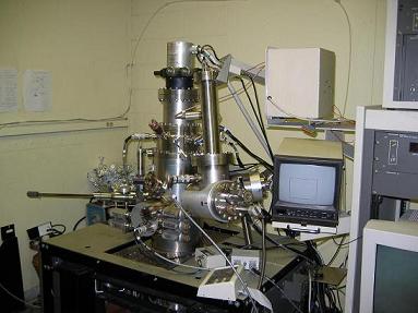
UHV - Ambient Pressure RAIRS System
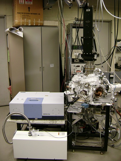
Our new high pressure RAIRS system contains two separate
chambers for low and high pressure analysis. The main chamber is a
typical UHV analysis chamber equipped with a quadrupole mass spectrometer
(Prisma QMS200 from Pfeiffer), and a combined reverse-view low energy
electron diffraction (LEED)- Auger electron spectrometer (AES) system
from LK technologies (Series RVL2000). The two instruments share a
common electron gun that is retractable for better positioning in the
chamber. A CMA2000 electron energy analyzer is used for the AES
experiments.
The high pressure IR cell is located underneath the main
chamber. It is equipped with a Bruker Vertex 70v FTIR; when
performing a high pressure experiment, the crystal is passed through a
differentially pumped sliding seal system into the high pressure cell.
With the crystal at the IR focus, the sample probe seals off the upper
UHV chamber from the high pressure IR cell.
The low pressure in the main chamber is maintained by a
turbo pump (Pfeiffer PM P02825), a titanium sublimation pump (Duniway
TSP-275-00), and an ion pump (Varian 9192641M001).
RAIRS System
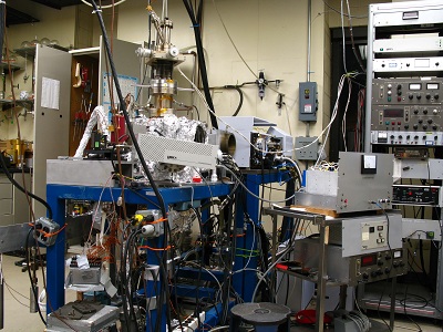
This chamber is equipped with an FTIR (Bruker
IFS-66v/s) for RAIRS studies, a quadrupole mass spectrometer (Hiden HAL
201/3F) interfaced to a personal computer for temperature programmed
desorption (TPD) studies, a single-pass cylindrical mirror
analyzer (Physical Electronics Model no. 10-155), and LEED
optics (Physical Electronics 15-180). It is pumped with a
turbo-molecular pump (Leybold-Heraeus TMP 360) backed by a
diffusion pump. It has all of the ancillary valves, vacuum
gauges, sample manipulator, gas doser, sputter ion gun, etc.,
needed for surface science studies of reactive chemistry on transition
metal surfaces.
UHV Scanning Probe Microscopy System

This variable temperature UHV Scanning Probe
Microscopy system was manufactured by Omicron Nanotechnology GmbH (http://www.omicron.de/). It was
purchased as a complete system and is equipped with a combined STM/AFM
unit with a sample stage that permits images to be obtained, in
principle, over the temperature range of 25 to 1500 K. The system
is also equipped with a reverse view LEED instrument and with a metal
evaporator. The STM uses a standard tube scanner with an
electrochemically sharpened metal tip whereas AFM images are obtained
with a needle-sensor based on a miniature quartz resonator. Sample
introduction and tip exchange are achieved without breaking vacuum
using a load lock system. The STM tip and AFM needle-sensor are
readily interchangeable. The capabilities of the STM function of
this instrument is demonstrated by images of the Si(111)-7x7 surface
obtained by our group.
RAIR-XPS System
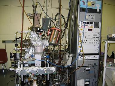
In addition to a dedicated FTIR (Mattson
RS-10,000) for RAIRS studies, this chamber is equipped with reverse-view
LEED optics (Princeton Research Instruments RVL 8-120), a
quadrupole mass spectrometer (UTI Instruments 100C) that is
fully computer controlled for multiplexed TPD studies, and a
hemispherical electron energy analyzer (VG Scientific CLAM 2)
and a dual Mg/Al anode X-ray source (VG Scientific) for XPS
studies. The chamber is pumped by a turbo-molecular pump
(Balzers TMP 520). The FTIR was purchased in 1984 from Mattson
and has been upgraded with new electronics and software and is
now controlled by a standard low cost personal computer. Although the
RAIRS-2 chamber is completely independent of the separately
described RAIRS-1 chamber, certain items such as IR detectors
and polarizers are shared, to some extent.
Scanning Tunneling Microscopy System
In addition to a UHV STM, this chamber is equipped with
reverse-view LEED optics (Princeton Research Instruments RVL
8-120), a q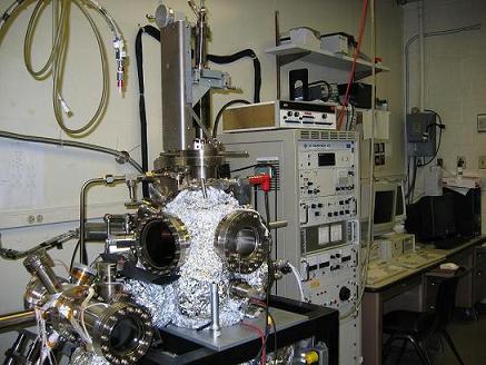 uadrupole mass spectrometer (UTI Instruments 100C) that
is fully computer controlled for multiplexed temperature programmed
desorption (TPD) studies, and a hemispherical electron energy
analyzer (VG Scientific CLAM 100) combined with a dual Mg/Al
anode X-ray source (VG Scientific) for XPS studies. Using an
electron gun the same analyzer is used for electron stimulated
Auger electron spectroscopy. The chamber is pumped by a
turbomolecular pump (Leybold Heraeus TMP 150), an ion pump
(Varian Associates, Inc., 230 l/s Starcell), and a Ti-sublimation
pump. The turbo pump can be isolated from the main chamber by a gate
valve and turned off during STM studies. The chamber pressure is
read with Bayard Alpert nude ion gauges (Granville Phillips Co.
series 274). The chamber rests on a vibrational isolation
platform (Barry Control, Inc.). The STM used in this study is a
UHV compatible commercial instrument (McAllister Technical
Services) employing a segmented piezo ceramic cylinder as the
scanning element. The one inch tube allows images of up to one
micron square to be obtained. The scanning tube is surrounded
by a larger piezo cylinder bonded to a quartz section, which in
turn supports two tungsten carbide rods on which the sample holder
sits. Sample approach to the tip is accomplished inertially under
computer control by applying a sawtooth wave-form to this larger
piezo tube. The chamber is configured for sample transfer via a
wobble stick between a fully rotatable XYZ manipulator and the
STM. The XYZ manipulator has a sample stage that can be cooled
with liquid nitrogen. The wobble stick also allows for transfer
of samples to and from a storage rack, which enablesup to six
samples to be present at once in the UHV chamber. The STM
contains a rack for storage of 8 tips, each of which can be
cleaned by heating within the UHV chamber by electron
bombardment. uadrupole mass spectrometer (UTI Instruments 100C) that
is fully computer controlled for multiplexed temperature programmed
desorption (TPD) studies, and a hemispherical electron energy
analyzer (VG Scientific CLAM 100) combined with a dual Mg/Al
anode X-ray source (VG Scientific) for XPS studies. Using an
electron gun the same analyzer is used for electron stimulated
Auger electron spectroscopy. The chamber is pumped by a
turbomolecular pump (Leybold Heraeus TMP 150), an ion pump
(Varian Associates, Inc., 230 l/s Starcell), and a Ti-sublimation
pump. The turbo pump can be isolated from the main chamber by a gate
valve and turned off during STM studies. The chamber pressure is
read with Bayard Alpert nude ion gauges (Granville Phillips Co.
series 274). The chamber rests on a vibrational isolation
platform (Barry Control, Inc.). The STM used in this study is a
UHV compatible commercial instrument (McAllister Technical
Services) employing a segmented piezo ceramic cylinder as the
scanning element. The one inch tube allows images of up to one
micron square to be obtained. The scanning tube is surrounded
by a larger piezo cylinder bonded to a quartz section, which in
turn supports two tungsten carbide rods on which the sample holder
sits. Sample approach to the tip is accomplished inertially under
computer control by applying a sawtooth wave-form to this larger
piezo tube. The chamber is configured for sample transfer via a
wobble stick between a fully rotatable XYZ manipulator and the
STM. The XYZ manipulator has a sample stage that can be cooled
with liquid nitrogen. The wobble stick also allows for transfer
of samples to and from a storage rack, which enablesup to six
samples to be present at once in the UHV chamber. The STM
contains a rack for storage of 8 tips, each of which can be
cleaned by heating within the UHV chamber by electron
bombardment.
Scanning Auger Multiprobe System - For Sale -
Contact RBD Instruments or the Trenary Group
directly for Details
The Perkin-Elmer Scanning Auger Microprobe 600 is a
complete system containing several instruments and all of the pumps,
gauges, valves, etc., needed for independent operation. The main
instrument is a cylindrical mirror analyzer with coaxial electron gun.
The electron gun (equipped with a LaB6 cathode) can be
focused to a beam size of 350 Å at a beam current of 0.1-.01 nA. The
beam can be rastered over the sample with the electron energy analyzer
tuned to a particular element. In this way, one can obtain a
two-dimensional map of an elemental distribution across a surface. The
scanning capabilities of the beam can be used with a secondary
electron detector to obtain scanning electron microscope (SEM) images
of a surface. The system also contains an ion gun for Auger depth
profile measurements and a quadrupole mass spectrometer for secondary
ion mass spectroscopy (SIMS). This system was purchased by the Amoco
Research Center in Naperville, IL in 1985 and was donated UIC in 1995.
Since then, the instrument has been enhanced in various ways including
upgrades of the computer and computer interface.
|